Bursa Foot Surgery Recovery
Overview
Infracalcaneal bursitis can significantly affect a person?s quality of life and his or her ability to perform activities of daily living, due to pain and impaired gait. This foot health problem may be diagnosed in several ways, including by palpation, or light pressure applied to the affected area by a healthcare practitioner. If the heel pain has existed for a long time, X-ray imaging studies may reveal localized calcification in the infracalcaneal bursa, though this is not always the case. MRI images are sometimes used as a diagnostic tool for this health problem, though MRI studies are considered unnecessary for diagnosis in many cases.
Causes
For the most part, it is a genetic condition. You develop it by inheriting a foot type that is prone to the mechanical irritation that leads to the problem. If you have this particular foot type and wear shoes that rub and irritate the back of the heel bone (calcaneus) where the Achilles tendon attaches, then you are even more likely to develop this type of bursitis. The contributing factors include a supinated foot type, a heel bone with a prominence (or pointed shape that stick out toward the tendon) at the back, a tight Achilles tendon (referred to as Equinus by doctors), and a high arch. All of these factors simply make it more likely that the back of the heel will press against the shoe and rub. As the tendon gets rubbed the wrong way, the bursa starts to develop and fill with fluid. This response is really your body's way of protecting the tendon from damage, but it backfires. The friction from all that rubbing causes the bursa to become inflamed and swell. This causes the bump to become red, hot and stick out even further. Much like a sore thumb, it then gets irritated by even the softest shoes.
Symptoms
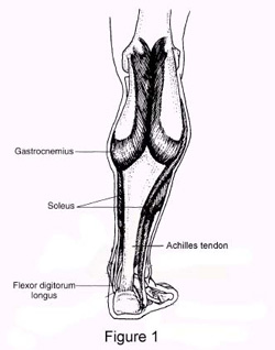
Below is a list of common signs and symptoms of retrocalcaneal bursitis. Recognizing and treating symptoms early can prevent retrocalcaneal bursitis from becoming chronic. Swelling. The retrocalcaneal bursa is located behind the Achilles tendon, just above where the tendon attaches to the heel bone. When the bursa is inflamed it will cause visible soft tissue swelling near the top of the heel bone. It is worth noting that bursitis of the retroachilles bursa, which is located between the Achilles tendon and skin, can manifest slightly differently: swelling may be more distinct, appearing as a hard lump behind the heel. Retroachilles bursitis is also more likely than retrocalcaneal bursitis to cause the skin at the back of the heel to turn red.
Diagnosis
Gram stain. A lab test called a Gram stain is used to determine if certain troublesome bacteria are present. Not all bacteria can be identified with a Gram stain, however, so even if the test comes back negative, septic bursitis cannot be completely ruled out. White blood cell count. An elevated number of white blood cells in the bursa's synovial fluid indicates an infection. Glucose levels test. Glucose levels that are significantly lower than normal may indicate infection.
Non Surgical Treatment
It is important to treat bursitis in the early stages to reduce the symptoms, minimize damage and maintain motion and strength in your foot. Resting your ankle, using proper cushioning, wearing comfortable footwear and reducing any activities that add pressure on your bursa will help to reduce your pain and bursitis inflammation.
Surgical Treatment
Surgery. Though rare, particularly challenging cases of retrocalcaneal bursitis might warrant a bursectomy, in which the troublesome bursa is removed from the back of the ankle. Surgery can be effective, but operating on this boney area can cause complications, such as trouble with skin healing at the incision site. In addition to removing the bursa, a doctor may use the surgery to treat another condition associated with the retrocalcaneal bursitis. For example, a surgeon may remove a sliver of bone from the back of the heel to alter foot mechanics and reduce future friction. Any bone spurs located where the Achilles attaches to the heel may also be removed. Regardless of the conservative treatment that is provided, it is important to wait until all pain and swelling around the back of the heel is gone before resuming activities. This may take several weeks. Once symptoms are gone, a patient may make a gradual return to his or her activity level before their bursitis symptoms began. Returning to activities that cause friction or stress on the bursa before it is healed will likely cause bursitis symptoms to flare up again.
Infracalcaneal bursitis can significantly affect a person?s quality of life and his or her ability to perform activities of daily living, due to pain and impaired gait. This foot health problem may be diagnosed in several ways, including by palpation, or light pressure applied to the affected area by a healthcare practitioner. If the heel pain has existed for a long time, X-ray imaging studies may reveal localized calcification in the infracalcaneal bursa, though this is not always the case. MRI images are sometimes used as a diagnostic tool for this health problem, though MRI studies are considered unnecessary for diagnosis in many cases.
Causes
For the most part, it is a genetic condition. You develop it by inheriting a foot type that is prone to the mechanical irritation that leads to the problem. If you have this particular foot type and wear shoes that rub and irritate the back of the heel bone (calcaneus) where the Achilles tendon attaches, then you are even more likely to develop this type of bursitis. The contributing factors include a supinated foot type, a heel bone with a prominence (or pointed shape that stick out toward the tendon) at the back, a tight Achilles tendon (referred to as Equinus by doctors), and a high arch. All of these factors simply make it more likely that the back of the heel will press against the shoe and rub. As the tendon gets rubbed the wrong way, the bursa starts to develop and fill with fluid. This response is really your body's way of protecting the tendon from damage, but it backfires. The friction from all that rubbing causes the bursa to become inflamed and swell. This causes the bump to become red, hot and stick out even further. Much like a sore thumb, it then gets irritated by even the softest shoes.
Symptoms
Below is a list of common signs and symptoms of retrocalcaneal bursitis. Recognizing and treating symptoms early can prevent retrocalcaneal bursitis from becoming chronic. Swelling. The retrocalcaneal bursa is located behind the Achilles tendon, just above where the tendon attaches to the heel bone. When the bursa is inflamed it will cause visible soft tissue swelling near the top of the heel bone. It is worth noting that bursitis of the retroachilles bursa, which is located between the Achilles tendon and skin, can manifest slightly differently: swelling may be more distinct, appearing as a hard lump behind the heel. Retroachilles bursitis is also more likely than retrocalcaneal bursitis to cause the skin at the back of the heel to turn red.
Diagnosis
Gram stain. A lab test called a Gram stain is used to determine if certain troublesome bacteria are present. Not all bacteria can be identified with a Gram stain, however, so even if the test comes back negative, septic bursitis cannot be completely ruled out. White blood cell count. An elevated number of white blood cells in the bursa's synovial fluid indicates an infection. Glucose levels test. Glucose levels that are significantly lower than normal may indicate infection.
Non Surgical Treatment
It is important to treat bursitis in the early stages to reduce the symptoms, minimize damage and maintain motion and strength in your foot. Resting your ankle, using proper cushioning, wearing comfortable footwear and reducing any activities that add pressure on your bursa will help to reduce your pain and bursitis inflammation.
Surgical Treatment
Surgery. Though rare, particularly challenging cases of retrocalcaneal bursitis might warrant a bursectomy, in which the troublesome bursa is removed from the back of the ankle. Surgery can be effective, but operating on this boney area can cause complications, such as trouble with skin healing at the incision site. In addition to removing the bursa, a doctor may use the surgery to treat another condition associated with the retrocalcaneal bursitis. For example, a surgeon may remove a sliver of bone from the back of the heel to alter foot mechanics and reduce future friction. Any bone spurs located where the Achilles attaches to the heel may also be removed. Regardless of the conservative treatment that is provided, it is important to wait until all pain and swelling around the back of the heel is gone before resuming activities. This may take several weeks. Once symptoms are gone, a patient may make a gradual return to his or her activity level before their bursitis symptoms began. Returning to activities that cause friction or stress on the bursa before it is healed will likely cause bursitis symptoms to flare up again.
Non Surgical Hammer Toe Treatments
 Overview
Hammer toes can result in pain and difficulty in moving the toe. Corns, calluses and blisters can occur from the rubbing of the contracted toe against the inside of the footwear. Both Hammer toe and mallet toe can cause pain during walking, running and other activities. If left untreated, the tendons of the toe may contract and tighten, causing the toe to become permanently stiff and contracted. A podiatric physician or surgeon may have to cut or realign tendons and/or remove pieces of bone to straighten the toe. This may require that the bones be fixed temporarily with pins while the toe heals.
Causes
Footwear can contribute significantly to the development of hammertoes. Shoes that are too small force your toes into a curled position. Over time, your toe tendons adjust to this positioning, causing your toe or toes to hold a hammered shape. Athletes may be especially susceptible, because of the increased forces on the toes from shoes that are too small or tight. Heel elevation in footwear is also problematic, as it causes your toes to be pushed into the shoe?s toe box. Heel elevation additionally contributes to muscle imbalance. A common example of this is when your Achilles tendon-the tendon at the back of your leg that attaches your calf muscles to your heel bone-is too tight, causing the tendons on the top of your foot that attach to your toes to work too hard and hold your toes in an unnatural, elevated position.
 Symptoms
Pain on the bottom of your foot, especially under the ball of your foot, is one of the most common symptoms associated with hammertoes. Other common signs and symptoms of hammertoes include pain at the top of your bent toe from footwear pressure. Corns on the top of your bent toe. Redness and swelling in your affected area. Decreased joint range of motion in your affected toe joints.
Diagnosis
Your healthcare provider will examine your foot, checking for redness, swelling, corns, and calluses. Your provider will also measure the flexibility of your toes and test how much feeling you Hammer toes have in your toes. You may have blood tests to check for arthritis, diabetes, and infection.
Non Surgical Treatment
If the affected toe is still flexible, you may be able to treat it by taping or splinting the toe to hold it straight. Your family doctor can show you how to do this. You may also try corrective footwear, corn pads and other devices to reduce pain. You may need to do certain exercises to keep your toe joints flexible. For example, you may need to move and stretch your toe gently with your hands. You can also exercise by picking things up with your toes. Small or soft objects, such as marbles or towels, work best. If your hammer toe becomes painful, you may need to apply an ice pack several times a day. This can help relieve the soreness and swelling. Nonsteroidal anti-inflammatory medicines (also called NSAIDs), such as ibuprofen (two brand names: Advil, Motrin) or naproxen (one brand name: Aleve), may be helpful. If your pain and swelling are severe, your doctor may need to give you a steroid injection in the toe joint.
Surgical Treatment
If your hammer, claw, or mallet toe gets worse, or if nonsurgical treatment does not help your pain, you may think about surgery. The type of surgery you choose depends on how severe your condition is and whether the toe joint is fixed (has no movement) or flexible (has some movement). A fixed toe joint often requires surgery to be straightened. A flexible toe joint can sometimes be straightened without surgery. Surgery choices include Phalangeal head resection (arthroplasty), in which the surgeon removes part of the toe bone. Joint fusion (arthrodesis), in which the surgeon removes part of the joint, letting the toe bones grow together (fuse). Cutting supporting tissue or moving tendons in the toe joint. How well surgery works depends on what type of surgery you have, how experienced your surgeon is, and how badly your toes are affected.
Physical Therapy After Bunion And Hammertoe Surgery
 Overview
A hammertoe occurs from a muscle and ligament imbalance around the toe joint which causes the middle joint of the toe to bend and become stuck in this position. The most common complaint with hammertoes is rubbing and irritation on the top of the bent toe. Toes that may curl rather than buckle, most commonly the baby toe, are also considered hammertoes. It can happen to any toe. Women are more likely to get pain associated with hammertoes than men because of shoe gear. Hammertoes can be a serious problem in people with diabetes or poor circulation. People with these conditions should see a doctor at the first sign of foot trouble.
Causes
Though hammer toes are principally hereditary, several other factors can contribute to the deformity. Most prevalent is an imbalance of the muscles and tendons that control the motion of the toe. When the tendon that pulls the toe upward is not as strong as the one that pulls it downward there is a disparity of power. This forces the toe to buckle and gradually become deformed. If the it persists, the toe can become rigid and harder to correct.
 Symptoms
For some people, a hammer toe is nothing more than an unsightly deformity that detracts from the appearance of the foot. However, discomfort may develop if a corn or callus develops on the end or top of the toe. If pressure and friction continue on the end or top of the toe, a painful ulcer may develop. Discomfort or pain can lead to difficulty walking.
Diagnosis
The treatment options vary with the type and severity of each hammer toe, although identifying the deformity early in its development is important to avoid surgery. Your podiatric physician will examine and X-ray the affected area and recommend a treatment plan specific to your condition.
Non Surgical Treatment
There is a variety of treatment options for hammertoe. The treatment your foot and ankle surgeon selects will depend upon the severity of your hammertoe and other factors. A number of non-surgical measures can be undertaken. Padding corns and calluses. Your foot and ankle surgeon can provide or prescribe pads designed to shield corns from irritation. If you want to try Hammer toes over-the-counter pads, avoid the medicated types. Medicated pads are generally not recommended because they may contain a small amount of acid that can be harmful. Consult your surgeon about this option. Changes in shoewear. Avoid shoes with pointed toes, shoes that are too short, or shoes with high heels, conditions that can force your toe against the front of the shoe. Instead, choose comfortable shoes with a deep, roomy toe box and heels no higher than two inches. Orthotic devices. A custom orthotic device placed in your shoe may help control the muscle/tendon imbalance. Injection therapy. Corticosteroid injections are sometimes used to ease pain and inflammation caused by hammertoe. Medications. Oral nonsteroidal anti-inflammatory drugs (NSAIDs), such as ibuprofen, may be recommended to reduce pain and inflammation. Splinting/strapping. Splints or small straps may be applied by the surgeon to realign the bent toe.
Surgical Treatment
Surgery to correct for a hammertoe may be performed as an outpatient procedure at a hospital, surgery center, or in the office of your podiatrist. There are multiple procedures that can be used depending on your individual foot structure and whether the deformity is flexible or rigid. There may be a surgical cut in the bone to get rid of an exostosis, or a joint may be completely removed to allow the toe to lay straight.
 Prevention
The easiest way to avoid hammertoe is to wear shoes that fit properly. Orthopaedic surgeons and podiatrists recommend shoes that have roomy toe boxes, which give the toes plenty of space to flex. Shoes that fit well should also cushion the arch in the middle of the foot. This helps to distribute the weight of the body evenly across the bones and joints of the foot. The size and shape of a foot can change with age, and many people inadvertently wear the wrong size shoe. Podiatrists recommend having your feet measured regularly to ensure that your shoes fit properly.
What Is Hallux Abducto Valgus?
Overview
 Bunion is a prominence of the medial portion of the head of the 1st metatarsal bone. The cause is often variations in position of the 1st metatarsal bone or great toe, such as lateral angulation of the great toe (hallux valgus). Secondary osteoarthritis and spur formation are common. Symptoms may include pain and redness, bursitis medial to the joint, and mild synovitis. Diagnosis is usually clinical. Treatment is usually a shoe with a wide toe box, protective pads, and orthotics. For bursitis or synovitis, corticosteroid injection may be helpful.
Causes
Many problems that occur in the feet are the result of abnormal pressure or rubbing. One way of understanding what happens in the foot due to abnormal pressure is to view the foot simply. Our simple model of a foot is made up of hard bone covered by soft tissue that we then put a shoe on top of. Most of the symptoms that develop over time are because the skin and soft tissue are caught between the hard bone on the inside and the hard shoe on the outside. Any prominence, or bump, in the bone will make the situation even worse over the bump. Skin responds to constant rubbing and pressure by forming a callus. The soft tissues underneath the skin respond to the constant pressure and rubbing by growing thicker. Both the thick callus and the thick soft tissues under the callus are irritated and painful. The answer to decreasing the pain is to remove the pressure. The pressure can be reduced from the outside by changing the pressure from the shoes. The pressure can be reduced from the inside by surgically removing any bony prominence.
Symptoms
Symptoms, which occur at the site of the bunion, may include pain or soreness, inflammation and redness, a burning sensation, possible numbness. Symptoms occur most often when wearing shoes that crowd the toes, such as shoes with a tight toe box or high heels. This may explain why women are more likely to have symptoms than men. In addition, spending long periods of time on your feet can aggravate the symptoms of bunions.
Diagnosis
A thorough medical history and physical exam by a physician is necessary for the proper diagnosis of bunions and other foot conditions. X-rays can help confirm the diagnosis by showing the bone displacement, joint swelling, and, in some cases, the overgrowth of bone that characterizes bunions. Doctors also will consider the possibility that the joint pain is caused by or complicated by Arthritis, which causes destruction of the cartilage of the joint. Gout, which causes the accumulation of uric acid crystals in the joint. Tiny fractures of a bone in the foot or stress fractures. Infection. Your doctor may order additional tests to rule out these possibilities.
Non Surgical Treatment
Treatment options vary with the type and severity of each bunion, although identifying the deformity early in its development is important in avoiding surgery. The primary goal of most early treatment options is to relieve pressure on the bunion and halt the progression of the joint deformity. A podiatrist may recommend these treatments. Padding and Taping, Often the first step in a treatment plan, padding the bunion minimizes pain and allows the patient to continue a normal, active life. Taping helps keep the foot in a normal position, thus reducing stress and pain. Medication, Anti-inflammatory drugs and cortisone injections are often prescribed to ease the acute pain and inflammation caused by joint deformities. Physical Therapy, Often used to provide relief of the inflammation and bunion pain. Ultrasound therapy is a popular technique for treating bunions and their associated soft tissue involvement. Orthotics, Shoe inserts may be useful in controlling foot function and may reduce symptoms and prevent worsening of the deformity. 
Surgical Treatment
If non-surgical treatments have failed to relieve your bunion pain, or when the pain is interfering with your daily activities, contact the Dallas bunion surgery specialists at North Texas Foot & Ankle to discuss surgical options. There are a several ways to perform bunion surgery. The best procedure for one person is not necessarily the best for another. Some procedures allow you to walk much sooner, reducing the need for crutches. Depending on your foot type, the procedure can have a greater risk for return of the bunion deformity. Other procedures may require you to be on crutches for a few weeks - but could offer a better result in the long-term. Learn more about the different types of bunion surgery.
Over-Pronation
Overview
Pronation is a normal motion which occurs after heel strike during walking. As the foot moves towards toe off, the foot naturally should move from an outside path towards the midline of the body stopping at neutral in ideal conditions. Overpronation is the foot?s motion past neutral and moving excessively toward the bodies midline. Overpronation can be caused by a number of factors including internal rotation of the femur or thigh bone, internal torsion of the tibia or shin bone, being genu valgum or ?knock kneed?, as well as fixed deformities of the ankle and forefoot. The overpronating foot is often hypermobile meaning it has excessive range of motion in the foot joints.
Causes
Generally fallen arches are a condition inherited from one or both parents. In addition, age, obesity, and pregnancy cause our arches to collapse. Being in a job that requires long hours of standing and/or walking (e.g. teaching, retail, hospitality, building etc) contributes to this condition, especially when standing on hard surfaces like concrete floors. Last, but not least unsupportive footwear makes our feet roll in more than they should.
Symptoms
Overpronation can lead to injuries and pain in the foot, ankle, knee, or hip. Overpronation puts extra stress on all the bones in the feet. The repeated stress on the knees, shins, thighs, and pelvis puts additional stress on the muscles, tendons, and ligaments of the lower leg. This can put the knee, hip, and back out of alignment, and it can become very painful.
Diagnosis
You can test for pronation by looking at the leg and foot from the back. Normally you can see the Achilles Tendon run straight down the leg into the heel. If the foot is pronated, the tendon will run straight down the leg, but when it lies on the heel it will twist outward. This makes the inner ankle bone much more prominent than the outer ankle bone.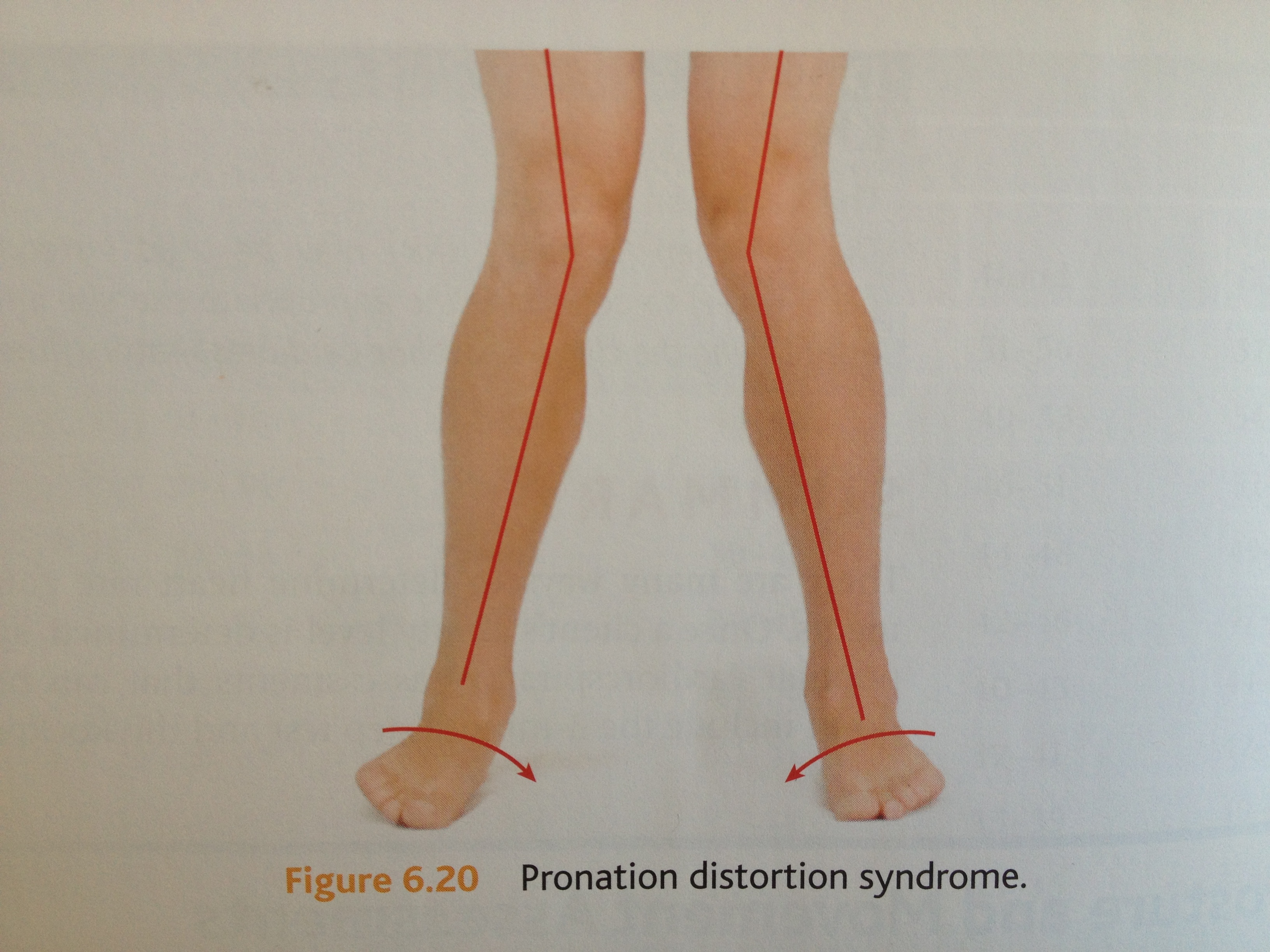
Non Surgical Treatment
Over-Pronation can be treated conservatively (non-surgical treatments) with over-the-counter orthotics. These orthotics should be designed with appropriate arch support and medial rearfoot posting to prevent the over-pronation. Footwear should also be examined to ensure there is a proper fit. Footwear with a firm heel counter is often recommended for extra support and stability. Improperly fitting footwear can lead to additional foot problems.
Prevention
Exercises to strengthen and stretch supporting muscles will help to keep the bones in proper alignment. Duck stance: Stand with your heels together and feet turned out. Tighten the buttock muscles, slightly tilt your pelvis forwards and try to rotate your legs outwards. You should feel your arches rising while you do this exercise. Calf stretch: Stand facing a wall and place hands on it for support. Lean forwards until stretch is felt in the calves. Hold for 30 seconds. Bend at knees and hold for a further 30 seconds. Repeat 5 times. Golf ball: While drawing your toes upwards towards your shins, roll a golf ball under the foot between 30 and 60 seconds. If you find a painful point, keep rolling the ball on that spot for 10 seconds. Big toe push:
Stand with your ankles in a neutral position (without rolling the foot inwards). Push down with your big toe but do not let the ankle roll inwards or the arch collapse. Hold for 5 seconds. Repeat 10 times. Build up to longer times and fewer repetitions. Ankle strengthener: Place a ball between your foot and a wall. Sitting down and keeping your toes pointed upwards, press the outside of the foot against the ball, as though pushing it into the wall. Hold for 5 seconds and repeat 10 times. Arch strengthener: Stand on one foot on the floor. The movements needed to remain balanced will strengthen the arch. When you are able to balance for 30 seconds, start doing this exercise using a wobble board.
What Is Severs Disease?
Overview
Sever's disease, is a musculoskeletal condition occurring in adolescence that symptomatically manifests as posterior heel pain during ambulation. Often participation in physical activity is severely limited resulting in frustration for children and parents alike. Conservative treatment options have included rest, abstinence from athletic activity, heel lifts, foot orthotic devices, ice, and calf-stretching exercise. The authors are proposing arch taping as an additional viable treatment option for controlling heel pain during athletic and other weight-bearing activities in patients with Sever's disease.
Causes
Your child may have pain along the edges of one or both heels during exercise. The pain usually goes away with rest, but may be worse in the morning. Your child's heel may also be slightly swollen and warm. The heel pain may be worse when your child climbs steps or stands on tiptoe. It may cause your child to limp.
Symptoms
Pain in the bottom surface and at the back of the heel. Extreme pain when the child places their heel on the ground. The pain is aggravated when running or jumping on hard surfaces. The pain is reduced when the child walks or runs on their toes.
Diagnosis
Sever?s disease is diagnosed based on a doctor?s physical examination of the lower leg, ankle, and foot. If the diagnosis is in question, the doctor may order x-rays or an MRI to determine if there are other injuries that may be causing the heel pain.
Non Surgical Treatment
In general, the goals for treatment include reducing the localized areas of inflammation. We recommend that patients utilize Ibuprofen every six to eight hours as needed. Stretching exercises on a daily basis, as well as prior to activity is thought to be helpful as well. Following work-outs and increased activities, it may be helpful to apply ice over affected areas. Heel cups are also available to provide cushion in shoes. In addition, for more severe symptoms, it may be helpful to refrain from sports and/or immobilize the area for a few weeks to help reduce the inflammation.
Recovery
This condition is self limiting, it will go away when the two parts of bony growth join together, this is natural. Unfortunately, Sever's disease can be very painful and limit sport activity of the child while waiting for it to go away, so treatment is often advised to help relieve it. In a few cases of Sever's disease, the treatment is not successful and these children will be restricted in their activity levels until the two growth areas join, usually around the age of 16 years. There are no known long term complications associated with Sever's disease.
Causes Signs Or Symptoms And Treating Of An Achilles Tendon Rupture
Overview
 An Achilles tendon rupture, or tear, is a common condition. This typically occurs in the unconditioned individual who sustains the rupture while playing sports, or perhaps, from tripping. There is a vigorous contraction of the muscle and the tendon tears.
Causes
The tendon usually ruptures without any warning. It is most common in men between the ages of 40-50, who play sports intermittently, such as badminton and squash. There was probably some degeneration in the tendon before the rupture which may or may not have been causing symptoms.
Symptoms
Ankle pain and swelling or feeling like the ankle has ?given out? after falling or stumbling. A loud audible pop when the ankle is injured. Patients may have a history of prior ankle pain or Achilles tendonitis, and may be active in sports. Swelling, tenderness and possible discoloration or ecchymosis in the Achilles tendon region. Indentation above the injured tendon where the torn tendon may be present. Difficulty moving around or walking. Individual has difficulty or is unable to move their ankle with full range of motion. MRI can confirm disruption or tear in the tendon. Inability to lift the toes.
Diagnosis
A physician usually can make this diagnosis with a good physical examination and history. X-rays usually are not taken. A simple test of squeezing the calf muscles while lying on your stomach should indicate if the tendon is still connected (the foot should point). This test isolates the connection between the calf muscle and tendon and eliminates other tendons that may still allow weak movement. A word of caution, Achilles tendon rupture is often misdiagnosed as a strain or minor tendon injury. Swelling and the continuing ability to weakly point your toes can confuse the diagnosis. Ultrasound and MRI are tests that can assist in difficult diagnosis. Depending on the degree of injury, these tests can also assist in determining which treatment may be best.
Non Surgical Treatment
Pain medicines can help decrease pain and swelling. A cast may be needed for 2 months or more. Your foot will be positioned in the cast with your toes pointing slightly down. Your caregiver will change your cast and your foot position several times while the tendon heals. Do not move or put weight on your foot until your caregiver tells you it is okay. A leg brace or splint may be needed to help keep your foot from moving while your tendon heals. Heel lifts are wedges put into your shoe or cast. Heel lifts help decrease pressure and keep your foot in the best position for your tendon to heal. Surgery may be needed if other treatments do not work. The edges of your tendon may need to be stitched back together. You may need a graft to patch the tear. A graft is a piece of another tendon or artificial material. 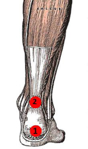
Surgical Treatment
Most published reports on surgical treatment fall into 3 different surgical approach categories that include the following: direct open, minimally invasive, and percutaneous. In multiple studies surgical treatment has demonstrated a lower rate of re-rupture compared to nonoperative treatment, but surgical treatment is associated with a higher rate of wound healing problems, infection, postoperative pain, adhesions, and nerve damage. Most commonly the direct open approach involves a 10- to 18-cm posteromedial incision. The minimally invasive approach has a 3- to 10-cm incision, and the percutaneous approach involves repairing the tendon through multiple small incisions. As with nonsurgical treatment there exists wide variation in the reported literature regarding postoperative treatment protocols. Multiple comparative studies have been published comparing different surgical approaches, repair methods, or postoperative treatment protocols.